loose body histology
Mucocele traumatized hemangioma pyogenic granuloma. A histopathological analysis of 119 surgically excised loose bodies revealed that the cases could be separated into three categories.

Qiao S Pathology Infarcted Appendix Epiploica Peritoneal Flickr
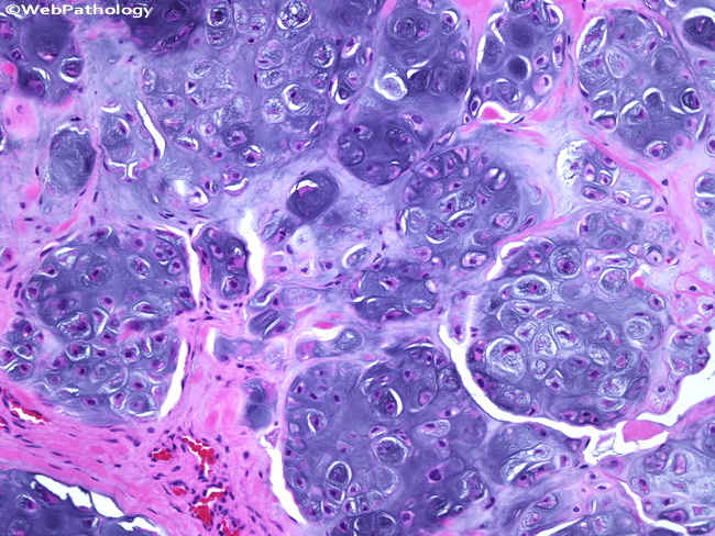
Synovial chondromatosis is a rare benign disorder of the synovium which leads to loose body formation due to metaplastic transformation.

. A study of loose bodies composed of cartilage or of cartilage and bone occuring in joints. We histologically examined 84 loose bodies and. Normally loose body is nourished by synovium and continues to.
Definition general Defined as infestation caused by body louse Pediculus humanus corporis Skin lesions due to direct bite hypersensitivity reaction and itching related. The loose bodies can vary in size from a few millimeters such as the size of a small pill to a few centimeters the size of a quarter. Blood vessel rich - key element proliferation of fibroblasts - key element inflammation - especially lymphocytes plasma cells common - evidence of erosionulceration.
Has the tide mark of articular cartilage has evidence of prior structure. Histologically based analyses of the nature and origin of loose bodies occurring in osteoarthrosis have been few and further study is warranted. The classification of loose bodies in human joints.
Synovial Chondromatosis is a proliferative disease of the synovium associated with cartilage metaplasia that results in multiple intra-articular loose bodies. The condition usually presents. The fragments can lead to damage to the articular cartilage.
Pathology Peritoneal loose bodies are formed by the torsion and autoamputation of epiploic appendages. As synovial membrane nodules were also classified to the same types as loose bodies. Loose bodies are usually asymptomatic 1.
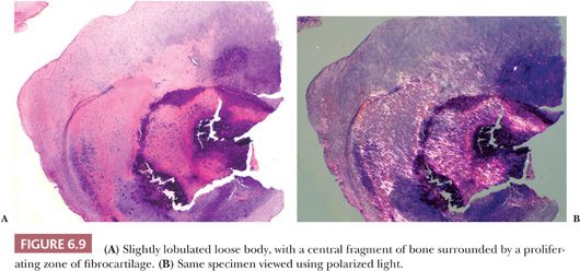
Histologic classification of loose bodies in osteoarthrosis. A rare pathology Abstract Loose bodies of the temporomandibular joint TMJ are an uncommon condition which can be caused by various complaints that can now be diagnosed with high. May form if portion of articular cartilage detached cartilage or cartilagebone within joint space with necrotic calcified centers may become attached to synovial membrane revascularize and convert to viable bone breaks off.
Peritoneal loose bodies are usually incidental findings at laparotomy. It presents as multiple cartilaginous. The loose bodies existed in the radiocarpal joint in 5 cases and all could be removed arthroscopically.
The various histologic characteristics of loose bodies in osteoarthrosis resulted from modifications including cartilage proliferation in the joint cavity and enchondral ossification in the synovial membrane. Two of the repaired tissues showed good histology after loose bodies that had been isolated in the joint for 12 weeks were fixed. In the other 5 cases the loose bodies were in the distal radioulnar joint and.
With special reference to their pathology and etiology A. Their sizes range from that of a pea 1-2 cm to giant loose bodies 5 cm or larger. Up to 10 cash back Histologically based analyses of the nature and origin of loose bodies occurring in osteoarthrosis have been few and further study is warranted.
Extracellular matrix was abundant. It is well known that loose bodies grow from proliferation of cartilage without blood supply in the joint cavity and that enchondral ossification is able to develop only under the condition of having a blood supply. BackgroundHistologically based analyses of the nature and origin of loose bodies occurring in osteoarthrosis have been few and further study is.
Histologically based analyses of the nature and origin of loose bodies occurring in osteoarthrosis have been few and further study is warranted.

Synovial Osteochondromatosis Of The Temporomandibular Joint A Case Report

Histological Slides Of Excised Synovial Chondromatosis Tissue At Power Download Scientific Diagram

A Rice Bodies Gross Specimen B Rice Bodies Microscopic Histology Download Scientific Diagram

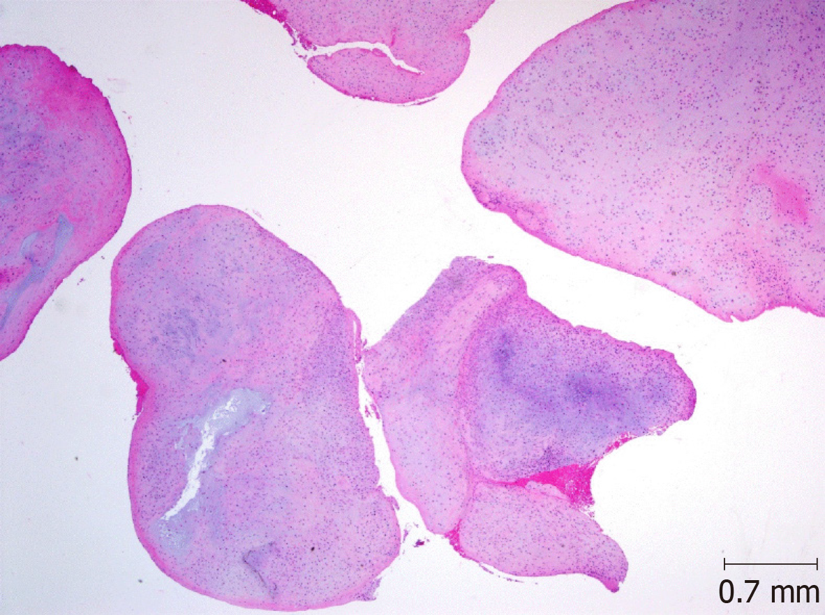
Histopathology Slide Of The Loose Body Showing Lobules Of Cartilage Download Scientific Diagram

Macro And Microscopic Findings Of The Loose Body A Extracted Loose Download Scientific Diagram

Figure 3 Arthroscopic Treatment Of A Case With Concomitant Subacromial And Subdeltoid Synovial Chondromatosis And Labrum Tear

Cartilage Proliferation Is Diagnosed During Histopathologic Evaluation Download Scientific Diagram

Qiao S Pathology Infarcted Appendix Epiploica Peritoneal Flickr

Qiao S Pathology Infarcted Appendix Epiploica Peritoneal Flickr

Pathology Outlines Synovial Tenosynovial Chondromatosis

Qiao S Pathology Infarcted Appendix Epiploica Peritoneal Flickr

Autoamputated Adnexa Presents As A Peritoneal Loose Body Fertility And Sterility

Pdf Uncalcified Synovial Chondromatosis In The Pisotriquetral Joint Semantic Scholar

Webpathology Com A Collection Of Surgical Pathology Images

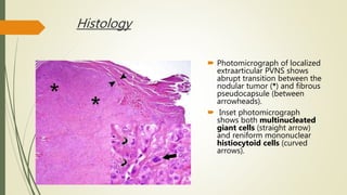
Pvns Synovial Chondromatosis Loose Bodies

Histologic Evaluation Of Osteochondral Loose Bodies And Repaired Tissues After Fixation Arthroscopy
Histological Examination Of The Loose Bodies Fibrous Connective Tissue Download Scientific Diagram


0 Response to "loose body histology"
Post a Comment